Knowledge Center · CBCT / 3D IMAGING
CBCT 3D Imaging: Turning Complex Diagnosis Into Shared Understanding
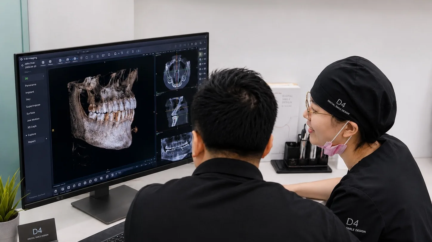
The value of CBCT is not only the 3D image itself. It helps patients, clinicians, and the treatment team discuss the same structural evidence.
When patients hear that a CBCT scan may be needed, the first reaction is often:
“Didn’t we already take an X-ray?”
That question is completely reasonable.
To patients, X-rays, panoramic images, and CBCT scans may all seem like “dental imaging.”
But clinically, they do not show the same information.
Two-dimensional images can tell us a lot: approximate tooth position, root condition, bone height, wisdom tooth direction, and some signs of pathology.
But they have one limitation.
They compress a three-dimensional structure into a flat image.
When the problem is simple, that may be enough.
But when treatment involves implants, root lesions, nerve canals, bone thickness, impacted teeth, complex root anatomy, or important anatomical structures, 2D imaging may not answer the key question.
In those situations, the value of CBCT is not “taking one more image.”
It is separating overlapping structures so they can be understood more clearly.
CBCT shows more than teeth
Many treatment risks are not on the visible tooth surface.
They are around roots, bone, nerves, sinuses, periapical tissues, and the spatial relationships between them.
For example, implant treatment is not simply placing an implant where a tooth is missing.
We need to understand bone height, bone width, nerve canal location, sinus distance, defect shape, and future prosthetic direction.
If these conditions are not understood, implant placement becomes an operation with unnecessary uncertainty.
CBCT helps reveal that uncertainty before treatment begins.
Why CBCT helps communication
Many clinical judgments are clear to the dentist but difficult for patients to imagine.
When a clinician says “there is not enough bone,” that can sound abstract.
When a clinician says “it is close to the nerve,” patients may understand there is risk but not where that risk is.
With CBCT, the clinician can review the image with the patient:
Here is the root.
Here is the bone boundary.
Here is the nerve canal.
Here is the lesion.
Here is the space that future implant or restoration must respect.
An abstract risk becomes a visible structure.
This does not mean the patient becomes the diagnostician.
It means the patient can understand why the clinician is making a particular decision.
Not everyone needs CBCT
CBCT is valuable, but not every patient needs it.
If clinical examination and 2D imaging already answer the question, there is no need to add an advanced scan just because it is available.
CBCT is usually needed when 2D imaging cannot answer the key question, treatment is close to important anatomical structures, or planning needs more precise spatial information.
The key is not the device itself.
The key is whether three-dimensional information is needed to reduce uncertainty.
How D4 uses CBCT
At D4, CBCT is not treated as a stand-alone imaging product.
It is one piece of structural evidence within treatment planning.
We look at CBCT together with intraoral scans, photos, X-rays, occlusal records, and clinical findings.
CBCT shows bone and internal structures.
Digital scans show tooth surfaces and occlusal relationships.
Photos show the smile, face, and aesthetic goal.
Clinical examination shows gum condition, mobility, pain, and function.
When these records are interpreted together, planning becomes more complete.
The point is not more data
Digital treatment is not about collecting more files.
If records are not interpreted properly, they simply become data storage.
The real value of CBCT is helping clinicians answer important questions before treatment begins:
Can this be done?
Where is the risk?
What needs to be addressed first?
Is there a more conservative path?
Does the patient understand the conditions behind this choice?
When these questions are clear, treatment does not begin with a procedure.
It begins with shared understanding.
The meaning of CBCT is not only seeing in three dimensions.
It is turning complex structural problems into a shared image that the clinician, patient, and team can discuss.
FAQ
How is dental CBCT different from a regular dental X-ray?
Regular dental X-rays are usually two-dimensional images. They are useful for observing general tooth structure, decay, root-tip problems, or local periodontal conditions. CBCT is three-dimensional imaging, allowing dentists to review teeth, roots, bone volume, nerve canals, the maxillary sinus, and nearby structures from different angles. It is not meant to replace every dental X-ray, but it can help clinicians understand spatial relationships that 2D images may not show clearly.
When is a CBCT scan needed?
CBCT is commonly used for implant planning, complex wisdom tooth evaluation, difficult root canal cases, root resorption, periapical lesions, periodontal bone defect assessment, and selected orthodontic or restorative evaluations. Whether CBCT is needed should not depend on whether the equipment is available, but on whether 2D records are enough to support the diagnosis. If a regular examination already answers the clinical question, CBCT may not be necessary.
Does CBCT involve a lot of radiation?
CBCT does involve radiation, so it should not be used casually as a routine scan. D4 uses a Planmeca CBCT system, which allows clinicians to choose an appropriate field of view, resolution, and Planmeca Ultra Low Dose™ 3D imaging protocol based on the purpose of the scan. The point of Ultra Low Dose™ is not simply to make the dose as low as possible, but to reduce unnecessary exposure while still meeting diagnostic needs. The dentist will choose an imaging approach that is sufficient for diagnosis without excessive exposure.
Why is CBCT often needed before dental implant treatment?
Implant planning is not only about the missing tooth area. The dentist also needs to evaluate bone volume, bone width, bone height, nerve canal position, sinus distance, and surrounding anatomical structures. CBCT helps assess implant position, direction, and potential risks before treatment, making the plan clearer and reducing uncertainty compared with relying only on visual examination or 2D imaging.
After taking a CBCT scan, what does the dentist mainly look at?
The dentist is not only looking for whether something is wrong. The scan is reviewed together with the treatment goal, including teeth, roots, bone, nerves, the maxillary sinus, lesion extent, and neighboring structures. In digital dentistry, CBCT can also be combined with intraoral scans, photography, and design records to support a more complete treatment judgment rather than relying on a single image alone.